Yes. Ultrasound uses sound waves, not radiation. It is safe for all ages, including babies and expectant mothers.
Understanding Ultrasound Scanning

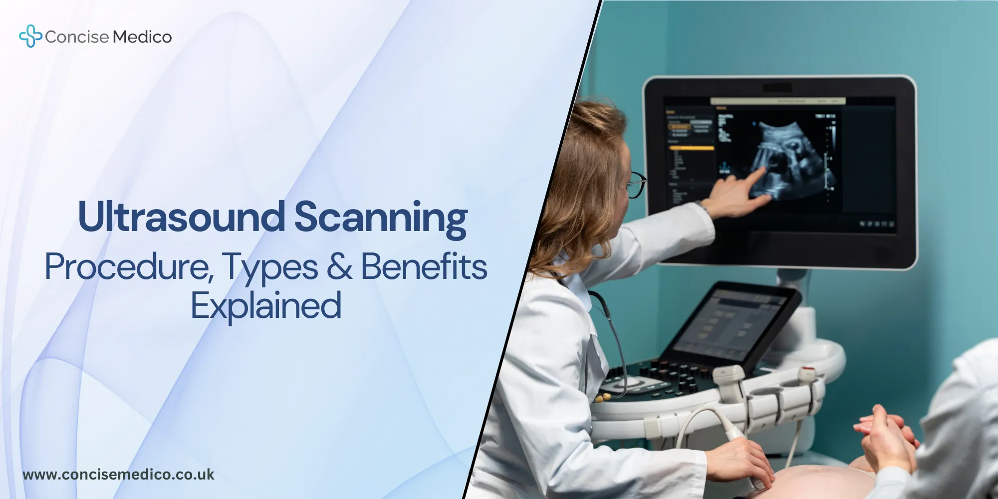
Understanding Ultrasound Scanning
Ultrasound technology uses high-pitched sound waves to take images inside the body. It is safe since it does not use ionising radiation. Ultrasound scanning is secure due to its lack of ionising radiation compared to X-rays. Doctors use ultrasound scanning to find problems early and to watch organ health.
In the UK, ultrasound is a trendy imaging method. The NHS says over 9 million scans happen each year. These scans help find health issues. Ultrasound imaging plays a main role in healthcare. More people should have access to it.
What is ultrasound technology?
It is the use of inaudible sound waves that bounce off tissues. A computer then turns those echoes into live images. This method is called ultrasound imaging technology. You feel no pain during a scan.
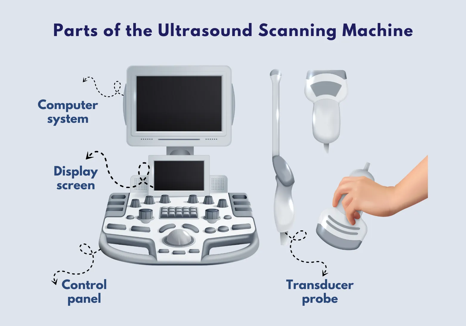
Parts of the Ultrasound Scanning Machine
-
Transducer probe
A hand-held tool that sends and receives sound waves. It comes in various shapes to scan different body parts and uses gel to improve contact.
-
Control panel
Lets the operator adjust scan depth, focus, and image presets. It has intuitive buttons and a touchscreen for quick changes during exams.
-
Display screen
Shows live, high-resolution pictures as the scan runs. Brightness and contrast controls help view details in dark or bright rooms.
-
Computer system
Converts echoes into clear images, stores scans in patient records, and runs advanced image-processing algorithms for better clarity.
The Science Behind Ultrasound Scanning Technology
A transducer sends short sound pulses into the body. These pulses hit organs or tissues and bounce back as echoes. The machine reads these echoes. It then draws them as images. You watch those images in real time.
Types of Ultrasound Scanning
-
2D ultrasound
A flat, black-and-white image used for basic checks. It shows a single slice of tissue at a time. Common in routine scans like gallbladder or thyroid exams.
-
3D ultrasound
Creates three-dimensional pictures that reveal shape and surface details. Doctors use it to assess fetal features or joint surfaces. It offers clearer views for planning treatments.
-
4D ultrasound
Provides live 3D video by adding motion over time. You can watch a baby move or a valve open and close in real time. It helps in detailed diagnostic and patient education.
-
Doppler ultrasound
Uses color to map blood flow speed and direction. It spots blockages, leaks, or vessel narrowing. It’s vital in heart exams, vascular checks, and stroke risk assessments.

-
Transvaginal ultrasound
This scan uses a small probe placed in the vagina to produce clear images of the pelvic organs. It shows the uterus, ovaries, and early pregnancy in good detail. Doctors use it for gynecological exams and to monitor fertility.
-
Endoscopic ultrasound
This scan attaches an ultrasound probe to an endoscope and guides it into the digestive tract. It gives clear views of the esophagus, stomach, pancreas, and nearby lymph nodes. Doctors use it to find gut problems and to guide biopsies.
Ultrasound Scanning Procedure: What to Expect
- It starts by applying clear gel to the skin. This helps sound waves travel.
- The probe moves slowly over the area.
- You watch live images on a screen.
- The scan takes about 15 to 45 minutes.
- You can leave right away—no recovery time.
Preparation and Aftercare
-
Before the scan
For pelvic exams, you may need a full bladder. Drink water one hour before. Wear loose clothes.
-
During the scan
Lie still and breathe normally. The nurse will guide you.
-
After the scan
Wipe off the gel. Drink, eat, and get back to your routine.
Advances in Ultrasound Scanning & Future Trends
Recent advances in ultrasound technology make scans clearer and faster. New tools include:
-
3D and 4D imaging
Gives depth views and live action videos. It helps with prenatal checks. It shows real-time movement of organs and fetuses. This helps doctors make precise diagnoses.
-
Contrast-enhanced ultrasound
Uses safe agents to highlight blood flow. It finds small tumours early. It can map tiny vessels that standard scans miss. This improves treatment planning for vascular diseases.
-
Portable devices
Handheld units connect to tablets or phones. They bring care to any location. They can run on battery power in remote areas. This expands access to urgent imaging outside hospitals.
-
AI-assisted analysis
Software that measures organs and spots issues in seconds. It learns from many scans to improve accuracy. It highlights areas of concern automatically. This speeds up reporting and reduces human error.
“In recent years, the integration of artificial intelligence (AI) has propelled ultrasound imaging to even greater heights. AI algorithms have shown remarkable capabilities in image analysis, aiding in tasks such as image segmentation, lesion detection, and tissue characterisation”
Karan Pandey, 203
-
Wearable ultrasound
Small patches that monitor organs all day. They send data to the cloud. They track changes over long periods without hospital visits. This enables continuous health monitoring at home.
-
Advanced contrast agents
Future agents will target specific tissues to spot disease early. They bind to markers on cells to highlight pathologies. They can reveal function as well as structure. This promises earlier detection of many conditions.
Why is Ultrasound Technology Important?
It spots diseases early. Early care leads to better outcomes. It monitors unborn babies safely. In emergencies, it reveals bleeding fast. Scans are quick, painless, and safe for all ages. New portable and wearable tools widen access. They cut travel and speed up care.
Advantages of Ultrasound Scanning
Key advantages of ultrasound include:
-
Safe and radiation-free
No harmful rays. Good for unborn babies and repeat exams. It poses no long-term health risks.
-
Real-time imaging
Live pictures guide instant decisions in surgery or injections. It lets doctors adjust procedures on the spot.
-
Cost-effective
It costs less than MRI or CT scans. Clinics can offer more scans at lower prices. It reduces patient bills.
-
Painless and non-invasive
No cuts, needles, or pain. You stay comfortable throughout. It reduces anxiety and stress.

-
Easily accessible
Found in most hospitals, clinics, and mobile units. It is quick to arrange. Patients wait less time for diagnosis.
-
Versatile
It scans soft tissues, blood flow, and moving organs. It serves many specialties. It adapts to each patient’s needs.
Learn more about ultrasound importance in legal settings.
Ultrasound Services We Offer
At Concise Medico, our ultrasound scanning services include:
-
Abdomen ultrasound
Detects stones, inflammation, and tumours. Fast and painless.
-
Hernia ultrasound
Finds hernias and measures their size for repair plans.
-
Kidney ultrasound
Checks for infections, stones, cysts, and tumours.
-
Testes ultrasound
Examines lumps and fertility issues with high detail.
Book Your Ultrasound Scan with Concise Medico
Our team uses top ultrasound technology for clear, fast results. Book your ultrasound scan today. Enjoy a comfortable experience with our caring staff. Contact us to set your appointment now.
FAQs

Questions about treatment?
Get in touch with our team for a free consultation and quote today.
Let’s TalkServices

Medical Reports
- Nationwide Medical Services.
- We provide fully compliant CPR reports.
- Psychologists, General Practitioners and Orthopaedics.

Rehabilitation
- Tailored Treatments for the Individuals.
- Assisting you on your road to Recovery.
- Access to over 1500 Certified Therapists.

Diagnostics
- Convenient locations.
- A wide range of Non-invasive Scans.
- Guaranteed Results within 28 days, no waiting list.
Personal Injury
- Nationwide Personal Injury Medical Assessments.
- Fully Compliant CPR Part 35 Expert Reports.
- Vetted Medical Experts Across All Injury Types.




We are medical expert witness reports providers with over a decade of experience in the medico-legal field. Our collective expertise counts for up to 45 years.
We give access to a nationwide network of medical expert witnesses and rehabilitation providers.
Services
Company
Resources
Open Hours
Monday to Friday
9am — 5pm


