TABLE OF CONTENT
MRI for Soft Tissue Injuries: What It Shows and When It’s Used
An NHS study of patients with soft tissue knee injuries found that around 40% of MRI scans confirmed ligament or meniscal injuries. This highlights the role of MRI in diagnosing joint and soft tissue damage.
Soft tissue injuries can affect your muscles, tendons, ligaments, cartilage and other tissues around the joints. These injuries may happen during the sport, exercise, work or everyday activities. Common signs include pain, swelling and limited movement. Doctors often recommend an MRI for soft tissue injury when they need a detailed look inside the body. Unlike an X-ray, an MRI can show soft tissues clearly. This helps doctors to find the exact cause of pain and plan the right treatment for you.
“MRI is one of the best tools we have for seeing soft tissue injuries in detail.”
– Dr Sarah Johnson, Consultant Radiologist
Role of MRI in Soft Tissue Injury
MRI plays a key role in soft tissue injury because it helps to find injuries that cannot be seen on an X-ray. It creates clear images of muscles, ligaments, tendons, cartilage and other soft tissues. An MRI scan for soft tissue injury helps doctors understand how serious an injury is and monitor the healing process over time. MRI does not use radiation; it is a safe imaging method for many patients when a detailed view of soft tissue damage is needed.
When Is an MRI Used For Soft Tissue Injuries?
An MRI for soft tissue injury is used when an X-ray or physical exam does not clearly show the actual problem. It helps find serious injuries such as torn ligaments, damaged tendons or injuries inside a joint, like an ACL or rotator cuff tear. An MRI scan for soft tissue injury gives a detailed view of the area which is injured. It helps doctors to see whether a muscle, tendon, ligament or cartilage has been affected.
Here are some conditions where an MRI is used for soft tissue injuries:
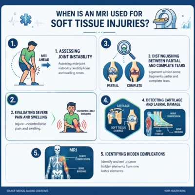
1. Assessing Joint Instability
Doctors may use an MRI for soft tissue injury if a joint feels loose, gives way or keeps moving out of place after an injury. An MRI can clearly show damaged ligaments, tendons and other soft tissues that help keep your joint stable. This helps doctors to find the cause of the problem and choose the right treatment.
2. Evaluating Severe Pain and Swelling
Severe pain and swelling that do not improve after a few days may mean there is more than a simple sprain or bruise. An MRI scan for soft tissue injury can be used to show the damage that cannot be seen on an X-ray, such as torn muscles, ligaments or tendons. It also helps doctors to understand how serious the injury is.
3. Distinguishing Between Partial and Complete Tears
An MRI helps doctors to see whether a ligament, tendon or muscle is partially torn or completely torn. This is important because a partial tear may heal with rest and therapy, while a complete tear may need surgery.
4. Detecting Cartilage and Labral Damage
Cartilage is the smooth tissue that cushions bones inside a joint, while the labrum is a ring of cartilage that helps to keep stable some joints, like the shoulder and hip. Damage to these tissues can cause pain, stiffness and clicking sounds. An MRI provides detailed images that help doctors to identify these injuries.
5. Identifying Hidden Complications
Some injuries are not easy to find during a physical exam or on an X-ray. An MRI for soft tissue injury can detect hidden problems such as deep muscle tears, tendon damage, trapped fluid or internal bleeding. Finding these issues early helps doctors to create the right treatment plan and reduce the risk of long-term problems.
What Can an MRI Show?
An MRI for soft tissue injury can show many types of damage. The scan produces detailed images of soft tissues and joints.
It may reveal:
- Ligament tears
- Tendon injuries
- Muscle strains
- Cartilage damage
Because the images are so detailed, doctors often identify the exact severity of the injury.
1. Ligament Tears
Ligaments connect bones to other bones and help keep your joints stable. A sudden twist or impact can cause a ligament tear. An MRI scan for soft tissue injury can help show whether the ligament is stretched, partially torn or completely torn. This is important when deciding whether surgery or physiotherapy is needed. Common ligament injuries include knee ligament tears, such as ACL injuries.
2. Tendon Injuries
Tendons connect muscles to bones. Overuse, sport or sudden strain can damage them. An MRI for soft tissue injury can help to show tendon inflammation, partial tears and complete tears. This helps doctors to understand the severity of the injury. Examples include Achilles tendon injuries and shoulder tendon tears.
3. Muscle Strains and Tears
Muscle injuries are common in sports and physical work. They can range from mild strains to complete tears. An MRI scan for soft tissue injury can help to show the size and location of the muscle damage. This helps estimate recovery time. The scan also helps detect bleeding and swelling within the muscle.
4. Cartilage Damage
Cartilage helps joints to move smoothly. Injury or wear and tear can damage it. An MRI can show the areas where cartilage has become worn, cracked or torn. This is especially useful in the knee, hip and shoulder. Finding cartilage damage early may help prevent further joint problems.

“Early diagnosis of ligament and tendon injuries can improve recovery and treatment planning.”
– Dr James Patel, Sports Medicine Specialist
Case Study: MRI Reveals Isolated Knee Ligament Tear
A 48-year-old woman developed severe pain, swelling and had difficulty putting weight on her left leg after a yoga session. Initial X-rays showed that there was no bone injury, but her symptoms suggested a soft tissue issue.
Doctors suggested an MRI scan for soft tissue injury, which revealed an isolated rupture of the anterolateral ligament (ALL) of the knee.
The MRI also confirmed that the meniscus, anterior cruciate ligament (ACL), posterior cruciate ligament (PCL) and collateral ligaments were intact, allowing doctors to find the exact injury.
Based on the MRI findings, she was treated without any surgery using rest, ice, anti-inflammatory medication and physiotherapy. The detailed MRI assessment helped her avoid unnecessary surgery.
Conclusion
Soft tissue injuries are not always easy to diagnose based on the symptoms alone. Pain, swelling and reduced movement can have many different causes. An MRI for soft tissue injury gives doctors a detailed view of muscles, ligaments, tendons and cartilage, helping them to identify the exact issue.
Whether your injury is caused by sport, work or everyday activities, early diagnosis can make a real difference. An MRI scan for soft tissue injury supports accurate treatment planning and may help reduce recovery time. At Concise Medico, we provide high-quality MRI services with fast appointments and detailed reporting. We help you from scheduling your appointment to the final MRI results.